Liver and pancreatic cancers are considered to be cancers that are easily detected late because early symptoms are not distinct. In particular, regarding the liver, in high-risk groups with chronic hepatitis or cirrhosis, it can be difficult to clearly distinguish the nature of the lesion using ultrasound alone, even if a small nodule develops. The pancreas is also located deep within the abdomen, which can limit its observation during standard abdominal ultrasound examinations.
For this reason, abdominal MRI is used as a precise evaluation test when liver or pancreatic disease is suspected or when ambiguous lesions are identified in conventional examinations. Since MRI allows for the visualization of minute tissue differences without radiation exposure, it helps to examine liver tumors, pancreatic lesions, and bile duct structures in greater detail.
Hepatocellular carcinoma, commonly referred to as 'liver cancer,' mostly occurs in high-risk groups such as those with hepatitis B or C and cirrhosis . Regular monitoring using liver ultrasound and serum alpha-fetoprotein (AFP) testing every six months is typically recommended for high-risk groups. However, additional detailed examinations, such as contrast-enhanced CT or MRI, may be performed if the lesion is unclear on ultrasound, if a small nodule is newly observed, or if there is a history of treatment for hepatocellular carcinoma requiring confirmation of recurrence.
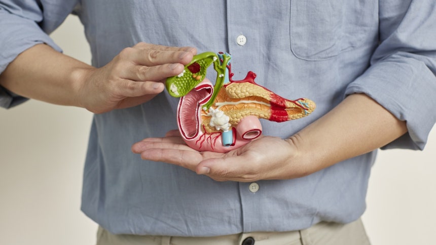
MRI scan capable of observing deep within the abdomen
In particular, MRI using gadocetic acid, a hepatocellular-specific contrast agent known as Primovist, is useful for determining the nature of small lesions as it allows for the simultaneous evaluation of changes in blood flow and hepatocellular function in liver nodules. Although it is sometimes difficult to confirm very small nodules less than 1 cm as cancer in a single MRI scan, tracking changes at regular intervals helps with early diagnosis and determining treatment strategies.
Precise imaging is crucial for pancreatic diseases , which are difficult to diagnose early . Since the pancreas is located in the retroperitoneum behind the stomach and intestines, it is easy to miss lesions in the body and tail during standard abdominal ultrasound examinations because they are obscured by intestinal gas or surrounding organs. Furthermore, early symptoms of pancreatic cancer are vague , so it is not uncommon for the disease to have already progressed by the time symptoms such as back pain, unexplained indigestion, weight loss, jaundice, or a sudden worsening of diabetes appear.
Kim Young-sun, Director of MRI Imaging at Mint Hospital (Radiology Specialist, Doctor of Medicine), explained , “Due to the anatomical location of the pancreas, it is sometimes difficult to fully confirm its entire structure through general health checkups or ultrasound examinations alone.” He added , “If pancreatic disease is suspected or there are risk factors such as a family history or pancreatic cysts, it is important to select precise examinations such as CT, MRI/MRCP, and endoscopic ultrasound according to the patient’s condition.”
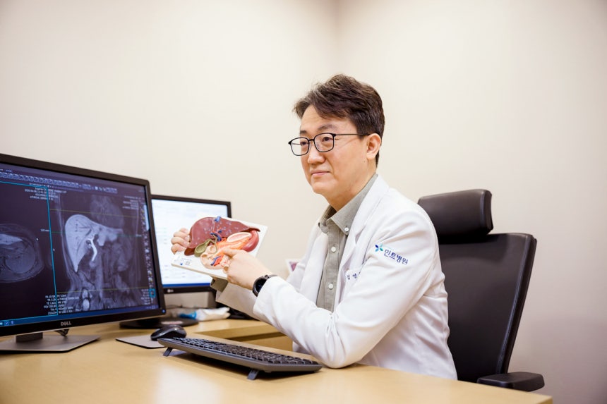
#Mint Hospital Director Kim Young-sun (Specializing in Hepatobiliary, Pancreatic, and Abdominal Surgery, Board Certified Radiologist/Doctor of Medicine)
MRI and MRCP allow for the simultaneous visualization of pancreatic parenchyma as well as pancreatic and bile duct structures, making them useful for evaluating pancreatic cystic lesions, bile duct dilation, and small pancreatic lesions. However, pancreatic MRI screening is not necessary for everyone. For the average risk group, it is advisable to consult with medical professionals to determine the necessity of the examination based on a comprehensive assessment of symptoms, family history, previous imaging results, and the pattern of diabetes development, rather than undergoing indiscriminate screening.
Recently, pancreatic screening MRI programs focusing on the pancreas and surrounding bile ducts are being introduced to reduce the cost burden of conventional abdominal MRI and to intensively examine necessary areas. This examination aims to more precisely examine the pancreas, which is difficult to detect during general health checkups, and can be selectively considered for high-risk groups or individuals suspected of having pancreatic disease.
Director Kim advised , “The earlier liver and pancreatic diseases are detected, the wider the treatment options become and the better the prognosis can be.” He added , “Just as important as precise imaging is maintaining proper abdominal breathing according to the medical staff’s instructions during the examination. Good respiratory cooperation is essential to reduce image distortion and obtain more accurate interpretation results.”
Reporter Jung Hee-won happy1@sportsworldi.com
https://n.news.naver.com/mnews/article/396/0000748155?sid=103