Women's Medical Center

4 Cases Where a Uterine-Pelvic MRI Is Absolutely Necessary

By 2026-09-08March 9th, 16No Comments


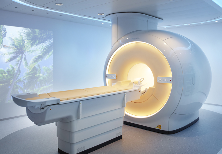
Ingenia 3T CX Mint Hospital MRI


In what situations is an MRI scan for uterine fibroids essential?


 MRI is an advanced diagnostic imaging technique that uses powerful magnetic fields and high frequencies to detect signals sent by water molecules within the body, creating three-dimensional images including horizontal, vertical, and cross-sectional views. It is primarily used for observing soft tissues, such as the nervous system, ligaments, and muscles, with great detail. 


In particular, uterine and pelvic MRI has excellent contrast (the ability to clearly distinguish objects), clearly distinguishing not only anatomical structures such as the uterus, ovaries, and uterine wall , but also tissue components such as blood, fat, and fluid, even without the use of contrast agents.


While ultrasound may not clearly show the ovaries and the back of the uterus depending on intestinal gas or location, the biggest advantage of MRI is that it allows for thorough examination without blind spots, regardless of location or the patient's body type .


Uterine Fibroid MRI, Essential When 1.

When you want treatment to preserve the uterus


I need treatment for unexplained menstrual cramps and irregular bleeding , but I'm worried that I might have to have my uterus removed.

This is the part that many people worry about the most after diagnosis. Fortunately, there are many methods available these days to treat only the fibroids while preserving the uterus without removing it. However, a pelvic MRI is essential for such delicate 'uterus-preserving treatment'.


While ultrasound results may vary slightly depending on the examiner's skill or equipment, MRI accurately displays the precise location of uterine fibroids and their relationship to surrounding organs without error. Based on this, it is possible to establish the optimal treatment direction and detailed plan to definitively resolve the fibroids while safely preserving your uterus.


Uterine Fibroid MRI, Essential When 2.

When assessing the suitability of a treatment method


The density of cells, blood flow, and water content of uterine fibroids vary widely from patient to patient. Furthermore, these minute tissue components can only be accurately identified through MRI .


Especially if you are considering non-surgical treatment, an MRI scan is a necessity, not an option. For example, if the cell density of the fibroid is too dense, the water content is excessive, or the blood flow is excessive, the efficiency of HIFU treatment may decrease.


By predicting this in advance through MRI results, we can quickly switch treatment methods to embolization or laparoscopic surgery instead of unnecessary procedures, thereby reducing the burden on the patient.


Uterine Fibroid MRI, Essential When 3.

When accurately evaluating post-treatment outcomes and the likelihood of recurrence


Uterine fibroids are also a disease prone to frequent recurrence . Ultrasound and CT scans have relatively low contrast, making it difficult to distinguish microscopic fibroids that remain as small as millet grains. On the other hand, MRI provides very clear images, allowing it to meticulously detect even minute uterine fibroid tissue that may have survived after treatment.


It is the most reliable test to definitively assess whether the treatment has been successfully completed and whether there is any possibility of recurrence that could cause menorrhagia or pelvic pain in the future.


Uterine Fibroid MRI, Essential When 4.

When a transvaginal ultrasound is burdensome or a malignant tumor is suspected
 

Minors who have never had sexual experience or unmarried women are prone to feeling significant fear and burden regarding the gynecological ultrasound examination itself, which involves inserting instruments into the vagina. While transrectal or abdominal ultrasounds are sometimes considered as alternatives, they often suffer from lower accuracy. In such cases, MRI serves as an excellent and comfortable alternative.


In addition , a detailed MRI examination plays a crucial role when malignancy (cancer) is suspected due to the irregular and unsettling shape of an ovarian mass (tumor) observed on ultrasound . Since the ovaries are hidden deep within the pelvis, pre-operative tissue biopsy is virtually impossible; therefore, MRI plays a decisive role in differentiating whether a lesion is benign or malignant before surgery.


​

250107 Uterine Fibroid MRI Card News 07


Mint Hospital

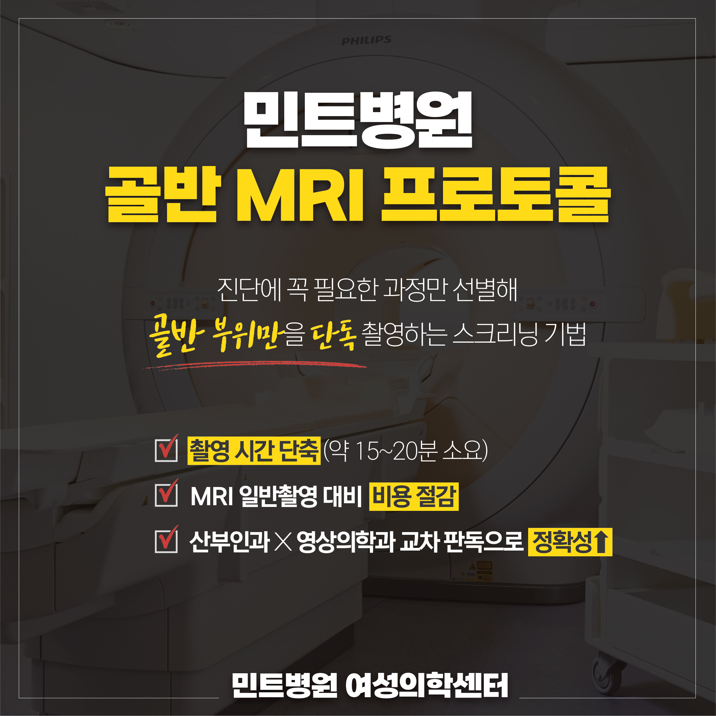
Pelvic MRI Examination Protocol

​

Select only the processes essential for diagnosis

Screening is performed exclusively on the pelvic area.

​


✅ Reduced shooting time

✅ Cost savings compared to standard MRI ⬇

✅ Increased accuracy through cross-reference of OB/GYN and Radiology

​


author avatar
Mint Hospital

Leave a comment